The eye is one of the most complex organs in the body. There are many parts that make up the eye and visual pathway, which allow us to experience vision.
Light rays enter the eye through the cornea, travelling through the small hole within the iris called the pupil. These light rays are focussed through the lens onto the retina, the tissue lining the back of the eye. The retina converts the light rays into electrical impulses, which are then sent through the optic nerve to the brain, where they are recognised as images.
Damage to any part of the eye or visual pathway to the brain can cause vision loss. In the case of inherited retinal diseases, the damage occurs in the retina, causing loss of the light-sensitive photoreceptor cells (rod and cones).
An image of the eye is detailed in the image below right, with the view from the top of the eyeball down. The components of the eye are described below.
Parts of the Eye
Cornea
The cornea is the transparent front layer of the eye that refracts, or redirects light to a sharp focus at the retina. Some inherited retinal diseases, such as Bietti’s crystalline dystrophy, can cause changes in the cornea, as well as in the retina.
Lens
The lens is a transparent structure inside the eye, that works with the cornea to focus the light. The lens in our eye is very similar in function to the lens of a camera, allowing us to change focus and hence see clearly at various distances.
A common, age-related condition that affects the lens is cataracts. Cataracts are very common in people over the age 60 years but can occur earlier in people with inherited retinal diseases. Cataracts are usually easily managed with cataract surgery, where the original lens is removed and replaced with a clear, artificial replacement lens.

Iris and Pupil
The iris is the visible layer, behind the cornea, that gives us our eye colour.
The iris is a muscle, that can adjust the size of the hole in its centre (the pupil) depending on the light levels in your surrounds. This means your pupil will be smaller in bright conditions to let less light in and bigger in dark conditions to let more light in.
In some inherited retinal diseases (such as achromatopsia), the pupil is slow to react to light changes, and people may be quite glare sensitive. In these cases, sunglasses can be very beneficial.
Vitreous
The clear, jelly-like substance found in the middle of the eye that helps to regulate eye pressure and shape.
Sclera
The sclera is the white outer coat of the eye. It is a tough, fibrous tissue, that protects the more fragile tissues inside the eyeball.
Optic Nerve
The optic nerve carries information about what you see from the retina to the brain.
When an ophthalmologist or optometrist looks into the eye, they can see the top of the nerve as it exits the eyeball, and can assess the health of the nerve.
Some inherited retinal diseases specifically target the optic nerve; for example, Leber hereditary optic neuropathy.
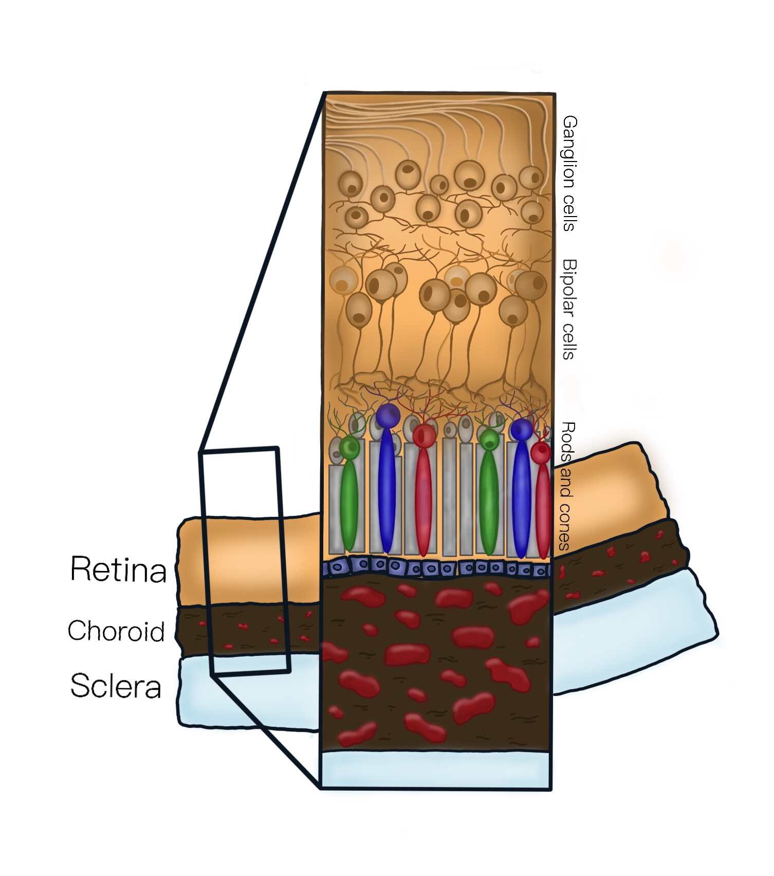
Retina
The retina is a light-sensitive tissue that lines the back of the eye. Although it is very thin and fragile (about the consistency of a wet piece of tissue paper), it contains several layers of cells and nerves.
The image on the left shows a cross-sectional representation of the retina showing, from top to bottom, the inner layers of the retina (inside the eyeball) through to the tough outer white layer of the eye (the sclera). The retina consists of different cell types including rod and cone photoreceptors, bipolar cells and ganglion cells. The retina is supplied with nutrients and oxygen from blood vessels in the choroid, which is the layer between the retina and sclera.
The layer of cells at the back of the retina called photoreceptors (rod and cone cells), are light-sensitive, and responsible for catching the light that is focused onto the retina.
The centre of the retina mostly has cone cells (which are responsible for daylight vision and colour vision), while the periphery of the retina has more rod cells (better at helping with night time vision).
Most inherited retinal diseases cause damage to the photoreceptors. The name of the disease can give an indication of the order of damage – cone-rod dystrophies damage the cone cells first (causing initial central vision loss), while rod-cone dystrophies damage the rod cells first (peripheral vision loss first).
Macula
The macula is the central part of the retina, that allows us to achieve high-resolution vision and accounts for our ability to read, recognise faces and to see the world in detail and colour.
A range of inherited retinal diseases can specifically target the macula, whilst not causing damage to the rest of the retina. These are called inherited maculopathies, and can sometimes be mistaken for the more common age-related macular degeneration.
Fovea
The fovea is the very centre of the macula, which provides our sharpest central vision. When you are asked to read letters on a vision chart at the eye doctors, this is a measurement of your foveal function.
Retinal Pigment Epithelium (RPE)
The retinal pigment epithelium is a single layer of cells located just behind the retina. The RPE provides a secure base for the photoreceptor cells of the retina to attach to, but is also responsible for providing nourishment to the retina from the blood supply on the other side (the choroid).
Choroid
The choroid is a layer containing blood vessels that bring oxygen and nutrients to the retina. The choroid lines the back of the eye, and is located between the retina and the sclera.
The inherited retinal disease choroideremia, starts in the choroid, and causes damage to the blood vessels, which then leads to death of the photoreceptor cells in the retina.